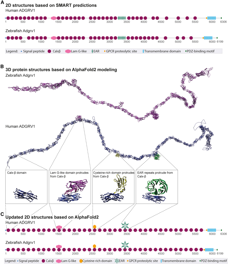
IMAGE

Figure 1

ID
ZDB-IMAGE-251004-87
Source
Figures for Stemerdink et al., 2025
Image
Figure Caption

Figure 1

In silico 3D structural modeling of the ADGRV1 protein identifies a previously undetected cysteine-rich domain and additional Calxβ domains compared to previous 2D-based models

(A) Schematic representation of the protein domain structure of the large isoform (isoform B) of human and zebrafish ADGRV1, based on 2D-SMART predictions. Both proteins exhibit a repetitive domain architecture, comprising a signal peptide, 35 Ca2+-binding calcium exchanger β domains (Calxβ), a laminin G-like domain (LamG-like), 6 epilepsy-associated repeats (EAR), a G-protein-coupled receptor (GPCR) proteolytic site, a seven-transmembrane region, and an intracellular region with a C-terminal class I PDZ-binding motif. Numbers indicate amino acid positions. (B) In silico 3D modeling of zebrafish (magenta) and human (blue) ADGRV1, based on AlphaFold2 predictions, suggests a more complex domain architecture than 2D-SMART-based models. 3D models revealed additional Calxβ domains as compared to the SMART-based predictions (39 instead of the previously predicted 35 Calxβ domains) and the presence of a previously unidentified cysteine-rich domain. Additionally, the 3D models reveal that the LamG-like domain, the cysteine-rich domain, and the EAR repeats protrude from the Calxβ domains, rather than existing as solitary domain structures that are separated by other (types of) domains. (C) Updated 2D schematic representation of human and zebrafish ADGRV1 (isoform B) based on novel 3D protein modeling.

Acknowledgments
This image is the copyrighted work of the attributed author or publisher, and ZFIN has permission only to display this image to its users. Additional permissions should be obtained from the applicable author or publisher of the image. Full text @ Mol Ther Nucleic Acids