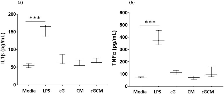
IMAGE

Figure 5.

ID
ZDB-IMAGE-251004-13
Source
Figures for Chugh et al., 2025
Image
Figure Caption

Figure 5.

Pro-inflammatory assessment of gelatin microspheres (cG), NK cell mimics (cGCM), NK cell membrane (CM) with differentiated THP-1 cells. Quantitative measurement of pro-inflammatory cytokines, (a) interleukin 1β (IL-1β), p< 0.0001, 95% CI [Media vs LPS] = −133.3 to −72.21, (b) tumour necrosis factor α (TNF-α) after 24 h incubation using enzyme-linked immunosorbent assay (ELISA), p < 0.0001, 95% CI [Media vs LPS] = −406.8 to −227.1, N = 3.

Lipopolysaccharides (LPS) was used as positive control. Graphs are plotted in box and whiskers format with max and min value showing all data points.

*p < 0.05. **p < 0.01. ***p < 0.001. ****p < 0.0001.

Acknowledgments
This image is the copyrighted work of the attributed author or publisher, and ZFIN has permission only to display this image to its users. Additional permissions should be obtained from the applicable author or publisher of the image. Full text @ J Tissue Eng