
|
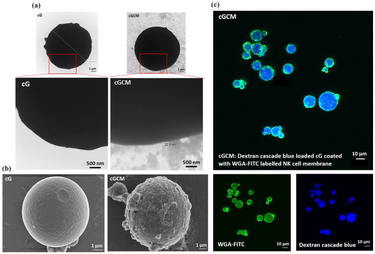
Figure 3.
Visualization of NK cell membrane onto gelatin microspheres: (a) transmission electron microscopy (TEM) images of uncoated gelatin microspheres (cG) and NK cell membrane-coated microspheres (cGCM). The cGCM images reveal a dark gelatin core with a surrounding lighter layer corresponding to the NK cell membrane coating. Due to the thin and low-contrast nature of the membrane layer, enlarged views of the red-highlighted regions are included to aid in visualizing coating integrity. Scale bar = 500 nm, (b) field emission scanning electron microscopic (FESEM) images of cG and cGCM, Scale bar = 1 μm, and (c) confocal laser scanning microscopic (CLSM) images of dextran cascade blue loaded gelatin spheres coated with WGA labelled KHYG-1 cell membrane, Scale bar = 10 μm.
Abbreviations: cG: cross-linked gelatin microspheres; cGCM: NK cell mimics, WGA-FITC: wheat germ agglutinin-fluorescein isothiocyanate.