IMAGE

Fig. 3

ID
ZDB-IMAGE-250927-16
Source
Figures for Wang et al., 2025
Image
Figure Caption

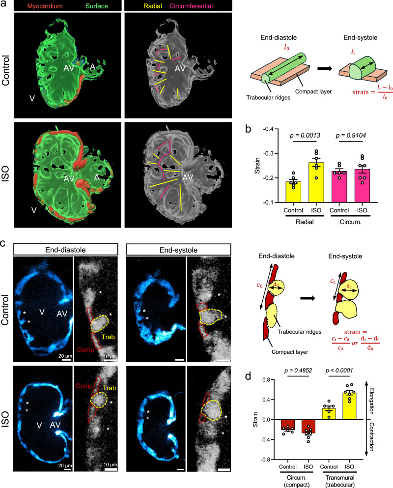
Fig. 3 ISO-mediated increase in strain aligning with radial trabecular ridges.

a, b 3-D volumetric and surface rendering revealed the network of radial (yellow) and circumferential (pink) trabecular ridges inside the hearts at 4 dpf. ISO treatment significantly increased the strain (i.e., shortening) along the radial trabecular ridges (n = 6 ridges for each group) but not the circumferential ridges (n = 6 ridges for control, n = 7 for ISO). c, d 2-D cross-sections of the trabeculae (asterisks) revealed that the surrounding compact myocardium (red dashed outlines) contracts circumferentially while the ridges (yellow dashed outlines) thicken (elongate) transmurally during systole. ISO treatment significantly increased the transmural strain within trabecular ridges (n = 6 ridges for control, n = 7 for ISO), whereas the circumferential strain in the surrounding compact layer remained similar (n = 6 ridges for control, n = 7 for ISO). Anatomic labels: V ventricle, A atrium, AV AV canal. All values are displayed with mean and standard error of mean (SEM). p-value is displayed for each comparison. One control heart and one ISO-treated heart were used for analysis. Ordinary one-way ANOVA followed by Šídák’s multiple comparisons test on the means was applied to determine statistical significance. Source data are provided as a Source Data file.

Acknowledgments
This image is the copyrighted work of the attributed author or publisher, and ZFIN has permission only to display this image to its users. Additional permissions should be obtained from the applicable author or publisher of the image. Full text @ Nat. Commun.