IMAGE

Figure 4

ID
ZDB-IMAGE-250924-53
Source
Figures for Kazzazy et al., 2025
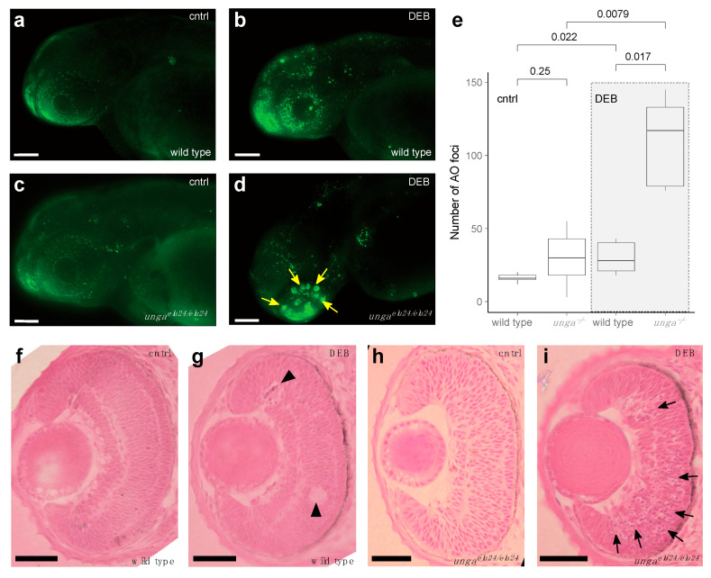
Image
Figure Caption

Figure 4

Sensitivity to DNA damage is increased in unga mutant zebrafish retinas. (a,b) AO staining of control and DEB-treated 2 dpf wild-type embryos shows an increased level of cell death upon the administration of the genotoxic agent. (c,d) DEB-treated unga mutant embryos also show elevated levels of apoptosis. However, unlike in wild-type controls, large clusters of AO-positive cells appear in these animals. Scale bar: 100 μm. (e) Quantification of apoptosis as measured by the number of AO foci in the control and DEB-treated embryos of the relevant genotypes (The Mann–Whitney test was used to calculate the p-values). (fi) HE staining of the retinal sections of the wild-type and unga mutant embryos reveal that upon DEB treatment, a few dying (or missing) cells can be seen in the wild-type animals (black arrowheads). In contrast, large clusters of cells with apoptotic character are apparent in the mutant retinas (black arrows). Scale bar: 100 μm.

Figure Data
Acknowledgments
This image is the copyrighted work of the attributed author or publisher, and ZFIN has permission only to display this image to its users. Additional permissions should be obtained from the applicable author or publisher of the image. Full text @ J Dev Biol