
Fig. 6
- ID
- ZDB-IMAGE-250917-61
- Publication
- Tevar et al., 2025 - Zebrafish adamtsl4 knockout recapitulates key features of human ADAMTSL4-related diseases: a gene involved in extracellular matrix organization, cell junctions and development
- All Figures
- Figures for Tevar et al., 2025
|
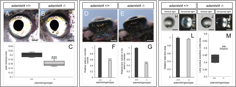
Fig. 6 Morphological phenotype characterization of 5 months F3 adamtsl4-KO zebrafish. A-B. Lateral morphology of the eye. (C) Quantification of pupil area relative to the eye area. Sample size: wild-type, n = 5; adamtsl4-KO, n = 9. D-E. Dorsal morphology of the eyes. F. Quantification of anterior chamber volume. G. Quantification of the visible lens volume within the anterior chamber. Sample size in F–G: wild-type, n = 20; adamtsl4-KO, n = 72. H-K. Lens morphology and light transmission. L. Quantification of total lens area. M. Quantification of lens nucleus area relative to the cortex area. Sample sizes: wild-type, n = 4; adamtsl4-KO, n = . ∗∗: p < 0.01; ∗∗∗: p < 0.001. Black asterisk: increased xanthophores. Black arrow: irregular pupil morphology. White asterisk: impaired light transmission in the lens. White arrow: direction of light. Cx, cortex; nu, nucleus.
Reprinted from Experimental Eye Research, , Tevar, A., Aroca-Aguilar, J.D., Atiénzar-Aroca, R., Ramírez, A.I., Fernández-Albarral, J.A., Escribano, J., Zebrafish adamtsl4 knockout recapitulates key features of human ADAMTSL4-related diseases: a gene involved in extracellular matrix organization, cell junctions and development, 110572, Copyright (2025) with permission from Elsevier. Full text @ Exp. Eye. Res.