
Fig. 5
- ID
- ZDB-IMAGE-250917-60
- Publication
- Tevar et al., 2025 - Zebrafish adamtsl4 knockout recapitulates key features of human ADAMTSL4-related diseases: a gene involved in extracellular matrix organization, cell junctions and development
- All Figures
- Figures for Tevar et al., 2025
|
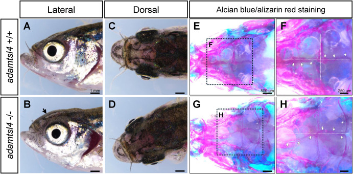
Fig. 5 Craniofacial characterization of 5 months F3 adamtsl4-KO zebrafish. A-D. Representative images of wild-type and adamtsl4-KO zebrafish before clearing and staining, highlighting gross craniofacial morphology. E-H. Staining with alcian blue for cartilage and alizarin red for bone structures reveals detailed craniofacial anatomy. Rectangles indicate regions of interest magnified in subsequent panels, providing a closer examination of structural differences between genotypes. White arrows indicate intracranial sutures. (For interpretation of the references to colour in this figure legend, the reader is referred to the Web version of this article.)
Reprinted from Experimental Eye Research, , Tevar, A., Aroca-Aguilar, J.D., Atiénzar-Aroca, R., Ramírez, A.I., Fernández-Albarral, J.A., Escribano, J., Zebrafish adamtsl4 knockout recapitulates key features of human ADAMTSL4-related diseases: a gene involved in extracellular matrix organization, cell junctions and development, 110572, Copyright (2025) with permission from Elsevier. Full text @ Exp. Eye. Res.