
Fig. 4
- ID
- ZDB-IMAGE-250917-59
- Publication
- Tevar et al., 2025 - Zebrafish adamtsl4 knockout recapitulates key features of human ADAMTSL4-related diseases: a gene involved in extracellular matrix organization, cell junctions and development
- All Figures
- Figures for Tevar et al., 2025
|
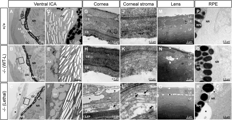
Fig. 4 Transmission electron microscopy of the ocular anterior segment in wild type and F3 adamtsl4 KO zebrafish larvae (6 dpf). Tissue sections were prepared as described in the Materials and Methods section. A-C. Representative photographs of wild type (+/+), wild type-like KO (−/− (WT-L)) and lethal KO (−/− (Lethal)) larvae. ICA: ventral iridocorneal angle. The rectangles indicate the areas magnified in subsequent panels. D-F. Ultrastructural details of iridophores and xantophores (XA), showing the presence of guanine crystals (GC) in the iridophores. G-I. Cornea. Note that the corneal endothelium was lost during tissue preparation in panels G and H. J-L. Ultrastructural details of the corneal stroma. M-O. Lens. P-R. Retinal pigment epithelium (RPE)-neuroretina (NR) interphase. CE, corneal epithelium; CEN, corneal endothelium; CF, collagen fibrils; CS, corneal stroma; GC, guanine crystals; LC, lens capsule; LEP, lens epithelium; LF, lens fibers; NR, neuroretina; RPE, retinal pigment epithelium; XA, xanthophores. Black arrows: enlarged and electron-dense CF. Black arrowhead: necrotic CE cell; black asterisks: abnormal intercellular separations; white arrowhead: autophagy; white arrows: thickened LC; white asterisks: swollen necrotic cells.
Reprinted from Experimental Eye Research, , Tevar, A., Aroca-Aguilar, J.D., Atiénzar-Aroca, R., Ramírez, A.I., Fernández-Albarral, J.A., Escribano, J., Zebrafish adamtsl4 knockout recapitulates key features of human ADAMTSL4-related diseases: a gene involved in extracellular matrix organization, cell junctions and development, 110572, Copyright (2025) with permission from Elsevier. Full text @ Exp. Eye. Res.