
|
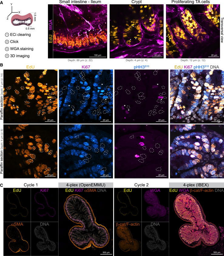
Figure 4
OpenEMMU for multiplexed imaging and DNA replication studies in the adult intestinal stem cell niche
(A) OpenEMMU compatibility with ethyl cinnamate tissue clearing and 3D confocal laser imaging, during analysis of DNA synthesis in the small intestine (ileum) of adult mice injected with EdU for 4 h. Several proliferating cell regions are observed, including the adult stem cell crypt and the transient amplifying (TA) cells in the villi, highlighted by the dotted line. Asterisks indicate mitotic cells.
(B) FFPE-processed ileum sections and confocal laser imaging of EdU in DNA-replicating cells, along with Ki67 and p-HH3S10 that are EdU positive, visualized by z stack confocal microscopy. Dotted strokes highlight EdU+ cells that are Ki67 negative. Asterisks indicate mitotic cells.
(C) FFPE-processed ileum section, visualized using tiling confocal microscopy. The section underwent two cycles of 4-plex staining, with OpenEMMU used in the first cycle and IBEX applied in the second (