
Fig. 3
- ID
- ZDB-IMAGE-250902-74
- Publication
- Abramova et al., 2025 - Disruption of grin2A, an epilepsy-associated gene, produces altered spontaneous swim behavior in zebrafish
- All Figures
- Figures for Abramova et al., 2025
|
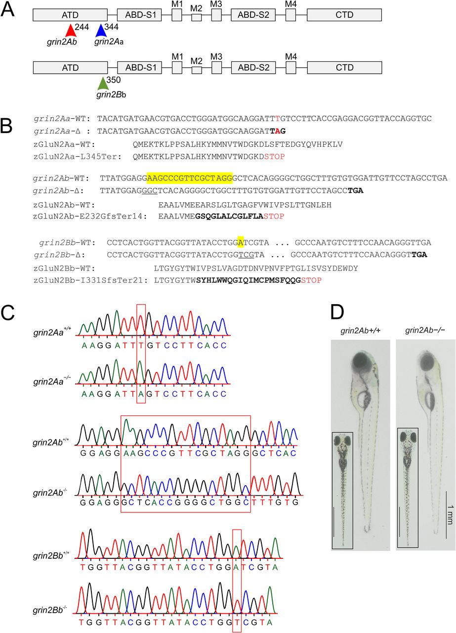
Fig. 3 Locations of zGluN2 mutations resulting in a premature stop codon and protein truncation. A, Schematic representation of the domain structure of a GluN2 subunit. It consists of four domains: the extracellular ATD and S1 and S2 lobes of the ABD; the membrane TMD with transmembrane segments M1, M3, M4, and M2 pore loop forming the ion channel; and the intracellular CTD. In the ATD, arrows indicate the position of grin2Aa mutation (sa14573) and gRNA target sites for grin2Ab and grin2Bb. B, The nucleotide and amino acid sequence alignment of the portion of the ATD of zGluN2Aa wild-type (grin2Aa-WT; zGluN2Aa-WT) and the mutant strain (grin2Aa-Δ; zGluN2Aa-L345Ter; top), zGluN2Ab wild-type (grin2Ab-WT; zGluN2Ab-WT) and the mutant strain (grin2Ab-Δ; zGluN2Ab-E232GfsTer14; middle), and zGluN2Bb wild-type (grin2Bb-WT; zGluN2Bb-WT) and the mutant strain (grin2Bb-Δ; zGluN2Bb-I331SfsTer21; bottom). C, Chromatograms of Sanger sequencing of PCR amplicons of genomic DNA from grin2Aa+/+ and grin2Aa−/− (top), grin2Ab+/+ and grin2Ab−/− (middle), and grin2Bb+/+ and grin2Bb−/− (bottom) larvae. D, Lateral and dorsal (inset) images of representative 6 dpf wild-type (grin2Ab+/+) and grin2Ab−/− larvae. For viability of larvae with mono- and biallelic deletion of grin2Aa, grin2Ab, or both genes at 6 dpf, see Extended Data Figure 3-1. For growth of the grin2Aa−/−, grin2Ab−/−, and grin2A−/− mutant fish during development, see Extended Data Figure 3-2.