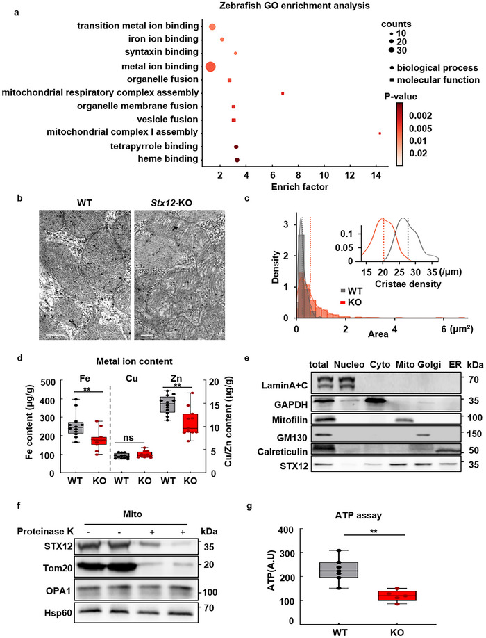
Abnormal mitochondrial morphology, ion imbalance, and energy deficiency in Stx12 KO mouse. a) Zebrafish RNAseq GO enrichment analysis demonstrated the association of Stx12 with iron binding, mitochondrial complex assembly, and vesicle fusion. b) Transmitting electron microscope images of heart sections in E18.5 wild type and Stx12‐KO mice. Representative EM image of heart section in Stx12‐KO (right), and wild type (WT) section (left). The scale bars represent 1 µm. c) Mitochondrial area and cristae density distribution of cardiac mitochondria. Mitochondria derived from Stx12‐KO (361 mitochondria from 3 KO) mice exhibited a significantly larger mitochondrial area than wild type (232 mitochondria from 2 WT). Inset showed that the cardiac mitochondria of Stx12‐KO (n = 57) had significantly decreased cristae density than the mitochondria of wild‐type (n = 57). Red dashed lines represented the averages (in mean ± SD) of Stx12‐KO mitochondrial area (0.61 ± 0.68 µm2) or cristae density (20 ± 3 µm−1), while gray dashed lines represented the mean of wild‐type mitochondrial area (0.28 ± 0.15 µm2, t‐test, p = 1.3 × 10⁻¹⁷) or cristae density (27 ± 3 µm−1, p = 1.7 × 10⁻²²). d) Metal ion content in the heart tissues of Stx12‐KO and wild‐type littermate mice. Scatterplots with boxplots showed that the iron and zinc quantity in the hearts of Stx12‐KO pups significantly decreased compared to wild types (WT) (Fe: WT, n = 12, 255.97 ± 66.44 ug g−1, KO, n = 11, 178.91 ± 52.02 µg g−1, p = 0.0017; Zn: WT, n = 12, 14.72 ± 1.98 ug g−1,KO, n = 11, 10.89 ± 3.07 µg g−1, p = 0.0017), while the copper quantity remained unchanged (Cu: WT, n = 12, 3.95 ± 0.66 ug g−1, KO, n = 12, 3.95 ± 0.66 µg g−1, p = 0.2456). e) Western blot analysis revealed STX12 broad distribution in mitochondria, Golgi, and ER in cardiac tissue. f) STX12 is located in the outer membrane of mitochondria. Mitochondria extracted from cardiac tissue were treated with proteinase K, which digested surface proteins of mitochondria. Tom20, OPA1, and Hsp60 are located on the outer membrane, inner membrane, and matrix of mitochondria, respectively. After protein K treatment, OPA1 and Hsp60 expression remained unchanged, while Tom20 and STX12 expression decreased, indicating that STX12 was located on the outer membrane of mitochondria. g) ATP assay of P0 mouse homogenized heart tissue. ATP assay of homogenized mouse heart tissue. Stx12 knock‐out cardiomyocyte exhibited a significantly decreased ATP level compared with normal mice. (WT, n = 7 mice; KO, n = 5 mice; p = 0.001183). Statistical results: **p < 0.01, ns, not significance; t‐test.
Acknowledgments
This image is the copyrighted work of the attributed author or publisher, and
ZFIN has permission only to display this image to its users.
Additional permissions should be obtained from the applicable author or publisher of the image.
Full text @ Adv Sci (Weinh)
Your Input Welcome
Thank you for submitting comments. Your input has been emailed to ZFIN curators who may contact you if
additional information is required.
Oops. Something went wrong. Please try again later.