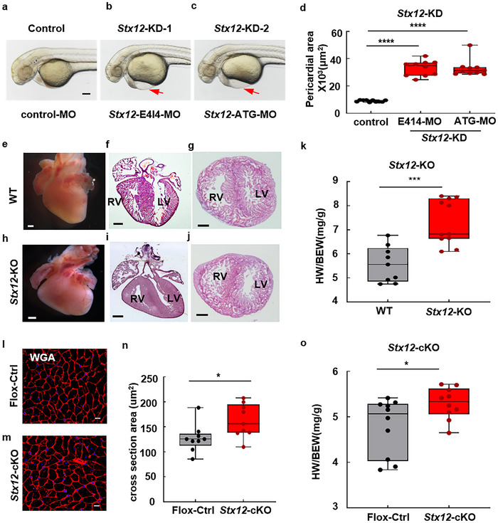
STX12 deficiency‐induced cardiac morphological changes in zebrafish and mice. a–c) Knockdown of Stx12 in zebrafish led to pericardial edema. The phenotypes of zebrafish were represented at 2 dpf after injection of morpholinos into zebrafish eggs to knock down STX12 expression. Scale bar, 100 µm. d) Quantification of pericardial area. The pericardial area of zebrafish significantly increased after Stx12 knockdown (control‐MO, n = 10, E414‐MO, n = 10, t‐test, p < 0.0001; ATG‐MO, n = 10, p < 0.0001). e,h) Cardiac stereomicroscopic images of P0 Stx12‐KO (e) and wild‐type littermates (h). Stx12‐KO heart exhibited cardiac malformation. Scale bar, 500 µm. f,g,i,j) Hematoxylin and eosin (HE) staining of coronal section (f, i) and cross section (g, j). Scale bars, 500 µm. k) Scatterplots with boxplots showed that heart weight‐to‐body weight ratio was significantly increased in Stx12‐KO mice (WT, n = 9, KO, n = 11, p = 0.0003). l,m) WGA staining of Stx12 myocardial‐specific knockout mice and Stx12‐flox control mice. Scale bar, 10 µm. n) Quantitative analysis of WGA staining (Flox‐control, n = 9, cKO, n = 9; p = 0.0272). o) Scatterplots with boxplots showed that the heart weight‐to‐body weight ratio was significantly increased in myocardial‐specific Stx12 knockout mice compared to Stx12‐flox control mice (Flox‐control, n = 10, cKO, n = 10, p = 0.0372). Statistical results: *p < 0.05, ***p < 0.001, ****p < 0.0001; t‐test.
This image is the copyrighted work of the attributed author or publisher, and
ZFIN has permission only to display this image to its users.
Additional permissions should be obtained from the applicable author or publisher of the image.
Full text @ Adv Sci (Weinh)
Your Input Welcome
Thank you for submitting comments. Your input has been emailed to ZFIN curators who may contact you if
additional information is required.
Oops. Something went wrong. Please try again later.