IMAGE

FIGURE 1

ID
ZDB-IMAGE-250828-104
Source
Figures for Malleret et al., 2025
Image
Figure Caption

FIGURE 1

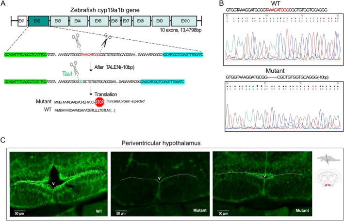
Cyp19a1b mutant zebrafish characterization. (A) The position of the 10 deleted nucleotides is shown on the top (red sequence), and the corresponding truncated protein is indicated below the gene sequence. The restriction enzyme cut site used for genotyping is written in green (TauI). Primer sequences used for genotyping are highlighted in green (forward) and blue (reverse). (B) Sequencing of the cyp19a1b gene in WT and mutant zebrafish showing the nucleotide deletion in exon 2. (C) Periventricular hypothalamus sections of AroB immunoreactivity in WT and mutant fish. Scale bars: 50 μm.

Acknowledgments
This image is the copyrighted work of the attributed author or publisher, and ZFIN has permission only to display this image to its users. Additional permissions should be obtained from the applicable author or publisher of the image. Full text @ J. Neurochem.