
Fig. 1
- ID
- ZDB-IMAGE-250820-12
- Publication
- Ameen et al., 2025 - Genomic Analysis of Glaucoma Pathogenesis Due to gmds Mutation in Zebrafish
- All Figures
- Figures for Ameen et al., 2025
|
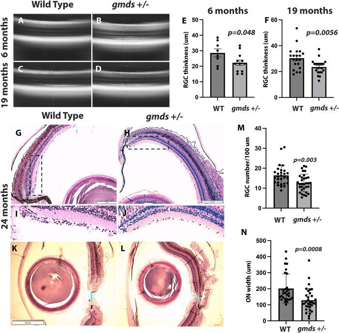
Fig. 1 Endophenotypes of glaucoma in heterozygous gmds mutant Zebrafish. Significantly thinner retinal ganglion cell (RGC) layers are found in gmds ± compared to wild-type siblings, as measured by SD-OCT imaging at 6 and 19 months; p = 0.048 and p = 0.0056, respectively) (A–F). H&E staining confirmed reduced RGC numbers in gmds mutants at 24 months; p = 0.003 (G-H, M). Boxed areas at the periphery of the retina are shown in (I and J). Significant optic nerve head width reduction p = 0.0008) is demonstrated in (K–N). Measurements were conducted in duplicate with 5 pairs of wild type and 5 pairs of mutant eyes per group.
Reprinted from Experimental Eye Research, , Ameen, M.T., Alloway, H., Longjohn, M.N., Gendron, R.L., Paradis, H., Benoukraf, T., French, C.R., Genomic Analysis of Glaucoma Pathogenesis Due to gmds Mutation in Zebrafish, 110497, Copyright (2025) with permission from Elsevier. Full text @ Exp. Eye. Res.