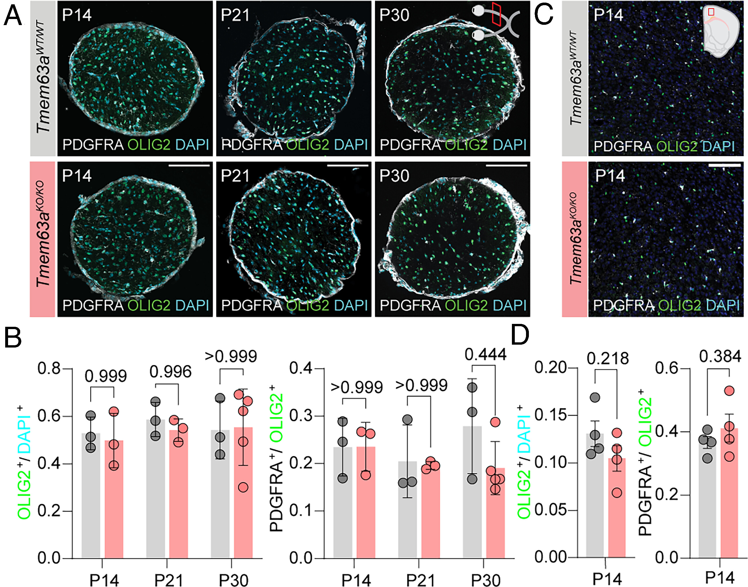
Fig. 5 Loss of Tmem63a does not affect OPC and OL populations. (A) Representative micrographs of 14 µm-thick optic nerve sections immunostained against OLIG2 (green) and PDGFRA (gray), and counterstained with DAPI (cyan) from Tmem63aWT/WT and Tmem63aKO/KO mice at P14, P21, and P30. (Scale bar, 100 µm.) (B) Left, percentage OLIG2+ cells/DAPI+ cells in the optic nerve for Tmem63aWT/WT and Tmem63aKO/KO mice. Right, percentage PDGFRA+ cells/OLIG2+ cells in the whole cross section of the optic nerve for Tmem63aWT/WT and Tmem63aKO/KO mice (n = 3 to 5 animals per genotype and age, Brown–Forsythe and Welch ANOVA). (C) Representative micrographs of 16 µm-thick cortical sections immunostained against OLIG2 (green) and PDGFRA (gray), and counterstained with DAPI (cyan) from Tmem63aWT/WT and Tmem63aKO/KO mice at P14. (Scale bar, 100 µm.) (D) Left, percentage OLIG2+ cells/DAPI+ cells in the cortex for Tmem63aWT/WT and Tmem63aKO/KO mice. Right, percentage PDGFRA+ cells/OLIG2+ cells in the cortex for Tmem63aWT/WT and Tmem63aKO/KO mice (n = 4 animals per genotype, unpaired t test).
Image
Figure Caption
Acknowledgments
This image is the copyrighted work of the attributed author or publisher, and
ZFIN has permission only to display this image to its users.
Additional permissions should be obtained from the applicable author or publisher of the image.
Full text @ Proc. Natl. Acad. Sci. USA

