
|
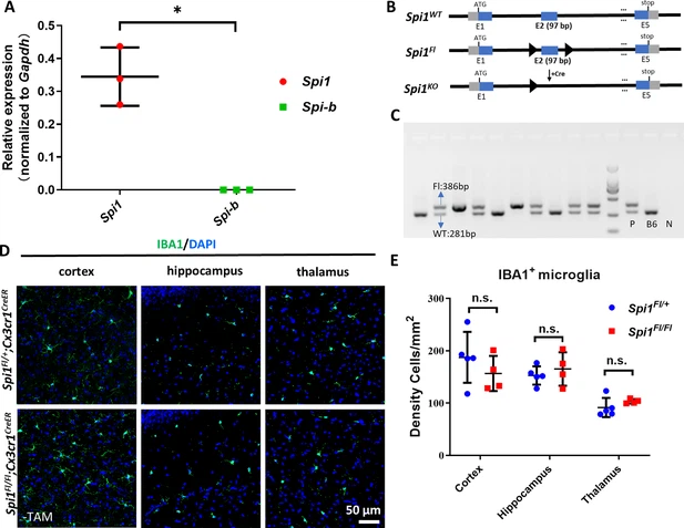
Fig. 6 - Supplemental 1 Adult microglia are not affected in Spi1Fl/Fl;Cx3cr1CreER mice. (A) Quantitative RT-PCR result shows the relative expression of Spi1 (n=3) and Spi-b (n=3) (normalized to Gapdh expression) in sorted microglia from adult mouse brain. (B) Schematic diagram shows the genomic loci of Spi1WT, Spi1Fl, and Spi1KO alleles, respectively. (C) Gel image shows the genotyping result of Spi1WT and Spi1Fl alleles (The image has been updated in the version of record). (D) Representative images of IBA1 and DAPI co-staining in the cortex, hippocampus, and thalamus regions of adult Spi1Fl+/;Cx3cr1CreER and Spi1Fl/ Fl;Cx3cr1CreERmice without tamoxifen (TAM) injection. (E) Quantification of the density (number) of IBA1+ microglia in the cortex, hippocampus, and thalamus regions of adult Spi1Fl/+;Cx3cr1CreER (n=5) and Spi1Fl/Fl;Cx3cr1CreER (n=4) mice. n.s.=not significant, p>0.05. *p<0.05.