
|
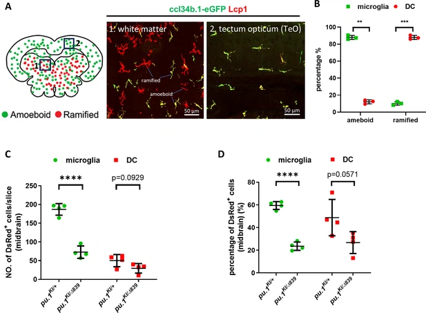
Fig. 3 - Supplemental 6 Conditional inactivation of Pu.1 leads to chronic elimination of microglia in the brain of adult zebrafish. (A) Representative images showing different morphology of microglia (ccl34b.1-eGFP+Lcp1+) and DCs (ccl34b.1-eGFP-Lcp1+) in two midbrain regions of TgBAC(ccl34b.1:eGFP) fish. The amoeboid and ramified cells were indicated accordingly. (B) Quantification of the proportion of microglia (ccl34b.1-eGFP+Lcp1+) (n=3) and DCs (n=3) (ccl34b.1-eGFP-Lcp1+) in total amoeboid and ramified Lcp1+ cells in the midbrain of TgBAC(ccl34b.1:eGFP) fish. (C) Quantification of the number of DsRed+ microglia and dendritic cells (DCs) on the midbrain cross section of pu.1KI/+;Tg(coro1a:CreER) (n=4) and pu.1KI/Δ839;Tg(coro1a:CreER) (n=4) fish at 3 mpi by amoeboid and ramified morphologies. (D) Quantification of the proportion of DsRed+ microglia and DCs on the midbrain cross section of pu.1KI/+;Tg(coro1a:CreER) (n=4) and pu.1KI/Δ839;Tg(coro1a:CreER) (n=4) fish at 3 mpi by amoeboid and ramified morphologies. **p<0.01; ***p<0.001;****p<0.0001.