
|
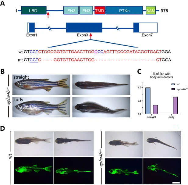
Fig. 2 - Supplemental 2 Zebrafish epha4b mutants exhibited body axis defects during development. (A) Diagram showing the protein domains, genomic structures, and sequences of the wild-type and corresponding epha4b mutants. Red arrows indicate mutation sites. (B) Representative images of epha4b mutants. (C) Bar graph showing the percentages of adult fish with normal and body axis defects in wild-type (n=39) and epha4b mutants (n=43). (D) Bright-field and GFP fluorescent images showing scoliosis epha4b mutants at 28 days post-fertilization (dpf) as indicated by Tg(Ola.Sp7:NLS-GFP) transgene, which labels the bone skeleton. Scale bars: 1 cm in panel (B), 2 mm in panel (D).