IMAGE

Figure 1

ID
ZDB-IMAGE-250728-18
Source
Figures for Boi et al., 2025
Image
Figure Caption

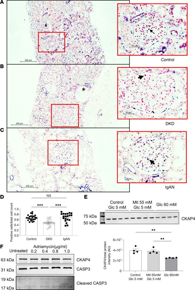
Figure 1 CKAP4 is downregulated in glomeruli in patients with DKD.

CKAP4 mRNA was detected using in situ hybridization in control (A), DKD patient biopsies (B), and IgAN patient biopsies (C). A purple/dark blue staining in the nuclear region characterizes cells positive for CKAP4 expression. Negative cells are characterized by a pink/red nuclear staining. The ratio of positive cells to total glomerular cells was used to quantify the extent of CKAP4 gene expression reduction in DKD (D). Completely differentiated human podocytes (HPODs) treated for 2 weeks with 60 mM glucose showed a 20%–30% reduction of CKAP4 (E). Treatment of HPODs with adriamycin for 24 hours does not cause a decrease in CKAP4 at protein level, although cleaved CASP3 level is increased in treated cells, indicating apoptosis (F). Unedited/uncropped total protein blots used for normalization calculation are provided as supplemental materials. Error bars represent average ± SEM. **P < 0.01, ***P < 0.001. D: 1-way ANOVA with Tukey’s multiple comparisons test, n = 25 (controls), 20 (DKD), 22 (IgAN) glomeruli. We scored 5 biopsies from 5 patients per group (4 for IgAN), and 5 (for controls, IgAN) or 4 (for DKD) glomeruli per biopsy. E: n = 4 replicates, 1-way ANOVA with multiple comparisons. The asterisk in B indicates a sclerotic glomerulus. The arrows in the respective zoomed-in sections of AC indicate cells showing positive staining for CKAP4. CASP3, caspase-3; CKAP4, cytoskeleton associated protein 4; DKD, diabetic kidney disease; Glc, glucose; IgAN, immunoglobulin A nephropathy, Mtl, mannitol.

Acknowledgments
This image is the copyrighted work of the attributed author or publisher, and ZFIN has permission only to display this image to its users. Additional permissions should be obtained from the applicable author or publisher of the image. Full text @ JCI Insight