
|
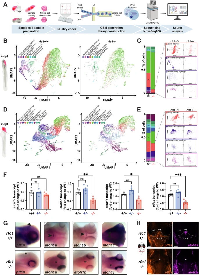
Fig. 4 RFC1 is expressed in early neural progenitors and is required for their proliferation.A Workflow for single-cell RNA sequencing (scRNA-seq). Microdissected heads (2 dpf) or brains (4 dpf) from rfc1+/+ and rfc1−/− larvae were pooled (three heads at 2 dpf or four brains at 4 dpf), dissociated, and processed for scRNA-seq using 10X Genomics technology. Created in BioRender. Samarut, E. (2025) https://BioRender.com/k7eukl7. B, D Uniform manifold approximation and projection (UMAP) plots of scRNA-seq data from 4 dpf (B) and 2 dpf (D) microdissected brains of rfc1+/+ and rfc1−/− larvae. Clusters of cells have been annotated following Seurat’s standard procedure, based on the accumulation of specific genes within top markers (see Supplementary Fig. S5B, C). C, E Proportions of cells in each identified cluster at 4 dpf (C) and 2 dpf (E), with UMAPs highlighting clusters significantly reduced in rfc1−/− brains. F RT-qPCR quantification of atoh1a, atoh1b, atoh1c, and ptf1a transcript levels in whole larvae at 3 dpf (rfc1+/+, rfc1+/–, and rfc1−/−, with n = 5 for each condition). G Whole-mount in situ hybridization for atoh1a, atoh1b, atoh1c, and ptf1a transcripts at 3 dpf in rfc1+/+ and rfc1−/− larvae. Expression in the cerebellum of wild-type larvae is indicated by arrowheads; loss of expression in mutants is marked by asterisks. H RNAscope in situ hybridization for ptf1a and atoh1a at 3 dpf in rfc1+/+ and rfc1−/− larvae. Scale bars: 50 μm. ot optic tectum, Va valvula cerebelli, CCe corpus cerebelli. Data in (F) are presented as mean ± SEM; individual dots represent biological replicates. Statistical analysis: one-way ANOVA with multiple comparisons. ns not significant (p > 0.05); * (p ≤ 0.05); ** (p ≤ 0.01); *** (p ≤ 0.001); **** (p ≤ 0.0001).