
Fig. 3
- ID
- ZDB-IMAGE-250724-56
- Publication
- Liu et al., 2025 - Identification and functional analysis of a novel TBC1D23 pathogenic variant in a Chinese family with pontocerebellar hypoplasia
- All Figures
- Figures for Liu et al., 2025
|
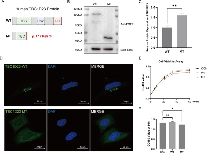
Fig. 3 The truncated TBC1D23 p.F171Qfs*8 protein is mislocalized, shows increased expression, and impairs cell proliferation. (A) Domain structure of wild-type (WT) and mutant (MT) TBC1D23. The p.F171Qfs*8 variant removes the Rhodanese and PH domains. (B, C) Western blot (B) and corresponding quantification (C) of EGFP-tagged proteins in HeLa cells show higher steady-state levels of the MT protein compared to WT. Data are mean ± SEM (n = 3); **P < 0.01 (Student’s t-test). (D) Confocal microscopy reveals normal Golgi localization for WT TBC1D23, whereas the MT protein is diffusely cytoplasmic. Green: EGFP-TBC1D23; Blue: DAPI. Scale bars, 20 μm. (E, F) CCK-8 assay (E) and endpoint analysis at 60 h (F) demonstrate that MT TBC1D23 expression significantly inhibits HeLa cell proliferation. Data are mean ± SEM (n = 3); *P < 0.05 (one-way ANOVA)