IMAGE

Fig 4

ID
ZDB-IMAGE-250722-37
Source
Figures for Kalifa et al., 2025
Image
Figure Caption

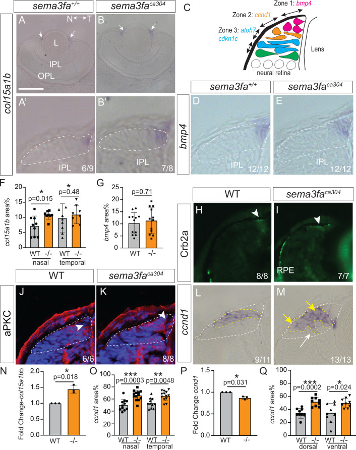
Fig 4 Disruption of retinal progenitor cell domain with loss of Sema3fa.

A-E) Zone 1 markers bmp4 (D, E) and col15a1b, at low (A, B) and high magnification (A′, B′), from plastic horizontal (A, B) and transverse (D, E) sections of WM ISH preparations. Schematic of the zonal structure of the CMZ (C). F-G) Average col15a1b (F; N = 2 independent experimental replicates) and dorsal bmp4 (G; N = 3 independent experimental replicates) expression domain areas normalized to CMZ area. H-M) Immunolabelling of distal CMZ cells for Crb2a/zs4 (H, I) and aPKC (J, K), and plastic sections of WM ccnd1 ISH (L, M). Arrowheads point to label (H, I, J, K) in the distal CMZ that contains stem cells. n’s represent numbers of embryos analysed that show WT-like expression (H-L), and dispersed ccnd1 expression (M; arrow). N) Fold change of col15a1b mRNA of sema3faca304 relative to WT 72 hpf eyes. Each dot is an independent biological replicate. Paired two-tailed t-test performed on delta Ct values. O) Average normalized ccnd1 ISH expression domain area for 72 hpf nasal/temporal CMZ in horizontal sections (O; N = 3 independent replicates). P) Fold change of ccnd1 (Q) mRNAs of sema3faca304 relative to WT 72 hpf eyes. Each dot is an independent biological replicate. Paired two-tailed t-test performed on delta Ct values. Q) Average normalized ccnd1 ISH expression domain area in 72 hpf dorsal CMZ in transverse sections (N = 3 independent replicates). For graphs in F, G, O, Q points are individual embryos, error is standard deviation, and statistics are Mann Whitney U-test. IPL, inner plexiform layer; L, lens; N, nasal; OPL, outer plexiform layer; RPE, retinal pigment epithelium; T, temporal. Scale bar in A is 100 µm for A, B; 30 µm for A′, B′, D, E, H-M.

Acknowledgments
This image is the copyrighted work of the attributed author or publisher, and ZFIN has permission only to display this image to its users. Additional permissions should be obtained from the applicable author or publisher of the image. Full text @ PLoS Genet.