IMAGE

Fig 2

ID
ZDB-IMAGE-250722-35
Source
Figures for Kalifa et al., 2025
Image
Figure Caption

Fig 2 
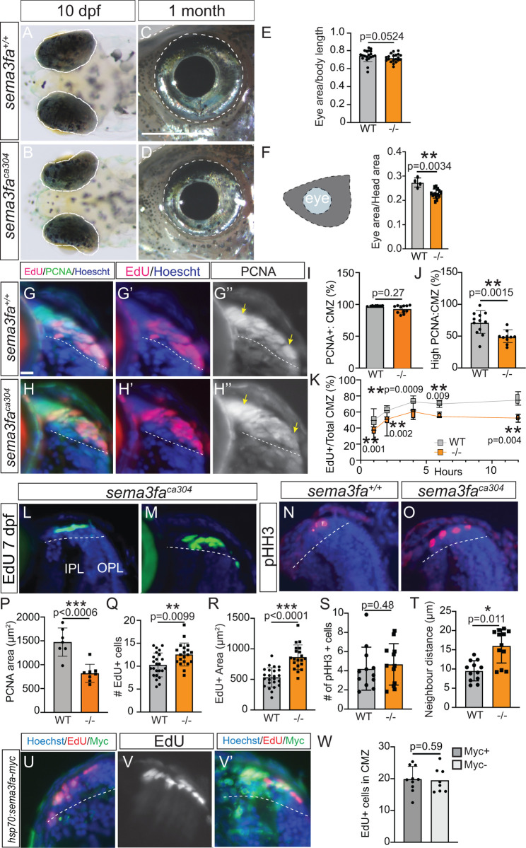
Larval eye size and CMZ proliferating progenitors impacted in the sema3faca304 mutant.

A-D) Dorsal view at 10 dpf (A, B) and lateral view at 1 month (C, D) of eyes (outlined in white dashed line) in WT (A, C) and sema3faca304 (B, D) fish. E-F) Eye area normalized to nose to swim bladder length at 10-11 dpf (E) and to area of the head (measured from the nasal-most aspect of the head to the anterior boundary of the gills) at 1 month (F) for WT and sema3faca304 fish. Pooled from N = 2 independent replicates. G-K) Cumulative EdU labelling of WT and sema3faca304 CMZ by bathing 72 hpf larvae in an EdU bath for 1-12 hours. One hour EdU labelling (G, G′, H, H′) of the CMZ in transverse retinal sections, also processed for PCNA immunolabelling (G, H, G′′, H′′) and for Hoescht to label nuclei (blue). The yellow arrows indicate uniform, intense PCNA immunolabel across the CMZ in WT (G′′) but lowered PCNA immunoreactivity in the mutant central CMZ (H′′). Graphs showing the percentage of Hoescht-labelled nuclei in the CMZ that are PCNA+ (I), the area of intense PCNA expression normalized to CMZ area (J), and the percentage of CMZ Hoescht-labelled cells that are EdU+ for each time point (K). Data pooled from N = 2 independent replicates. L-O) Retinal cryosections of 7 dpf CMZ showing EdU-labelled progenitors after a 4-hour EdU pulse (L, M) and immunolabelling for pHH3 (N, O). P-T) Graphs representing quantitation from retinal sections of the area of PCNA immunolabelling for the dorsal and ventral retina (P; N = 2 independent replicates), numbers of EdU+ cells (Q; N = 3 independent replicates) and the area of the CMZ they occupy (R; N = 3 independent replicates) from horizontal retinal sections, numbers of pHH3 + cells (S; N = 3) and the mean distance between them (T; N = 3) from horizontal retinal sections. U-W) Quantitation (W; N = 3 independent replicates) of the numbers of EdU+ progenitors (8 hour pulse) in Sema3fa-Myc-positive and Sema3fa-Myc-negative dorsal CMZ in transverse retinal sections of heat-shocked 5 dpf larvae injected at the one-cell stage with a construct with sema3fa-myc under the control of the hsp70 promoter. The ventral (U) and dorsal (V, V′) CMZ of a single retinal section from a heat-shocked larvae, showing little (U) or Myc (V′; green) immunolabelling for Sema3fa-myc+ along with Hoechst (blue) and EdU (U, V, V′; red) label. For all graphs, values represent individual embryos, error is standard deviation, and Mann Whitney-U test was used to test statistical significance. IPL, inner plexiform layer; OPL, outer plexiform layer. Scale bar in C represents 500 µm for A, B and 1000 µm for C, D; scale bar in G is 10 µm for G-H, L-O and U-V.

Acknowledgments
This image is the copyrighted work of the attributed author or publisher, and ZFIN has permission only to display this image to its users. Additional permissions should be obtained from the applicable author or publisher of the image. Full text @ PLoS Genet.