IMAGE

Fig 1

ID
ZDB-IMAGE-250722-33
Source
Figures for Kalifa et al., 2025
Image
Figure Caption

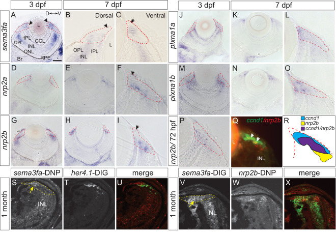
Fig 1 Expression of mRNAs for Sema3fa and its receptors in the larval and juvenile CMZ.

Plastic sections through the eyes of WM ISH of 3 (A, D, G, J, M, P) and 7 (B, C, E, F, H, I, K, L, N, O) dpf larvae for sema3fa (A-C) and potential receptors, nrp2a(D-F), nrp2b(G-I, P), plxna1a(J-L), and plxna1b(M-O). The CMZ (red dashed lines) is shown enlarged at 7 dpf (B, C, F, I, L, O). Peripheral-most CMZ lacks expression (arrows B, C, F, I). Q) Double FISH for nrp2b (red) and ccnd1 (green) mRNA in the 72 hpf CMZ. R) Schematic representing general organization of ccnd1 (blue) and nrp2b (yellow) expression in the CMZ. The two expression domains exhibit some overlap (purple). S-X) Double FISH on sections of 1-month retinas with a DNP riboprobe for sema3fa (S) and DIG probe for her4.1 (T; merge in U), and with a DIG probe for sema3fa (V) and DNP probe for nrp2b (W; merge in X). Expression of sema3fa in CMZ (dotted yellow line) indicated by yellow arrows. Orientation bar in A applies to A, D, E, G, H, J, K, M, N. D, dorsal; GCL, ganglion cell layer; INL, inner nuclear layer; IPL, inner plexiform layer; L, lens; ONL, outer nuclear layer; outer plexiform layer, OPL; RPE, retinal pigment epithelium; V, ventral. Scale bar in A is 50 µm for A, D, E, G, H, J, K, M, N and 10 µm for all other panels.

Acknowledgments
This image is the copyrighted work of the attributed author or publisher, and ZFIN has permission only to display this image to its users. Additional permissions should be obtained from the applicable author or publisher of the image. Full text @ PLoS Genet.