IMAGE

Fig. 4.

ID
ZDB-IMAGE-250722-18
Source
Figures for Feng et al., 2025
Image
Figure Caption

Fig. 4.

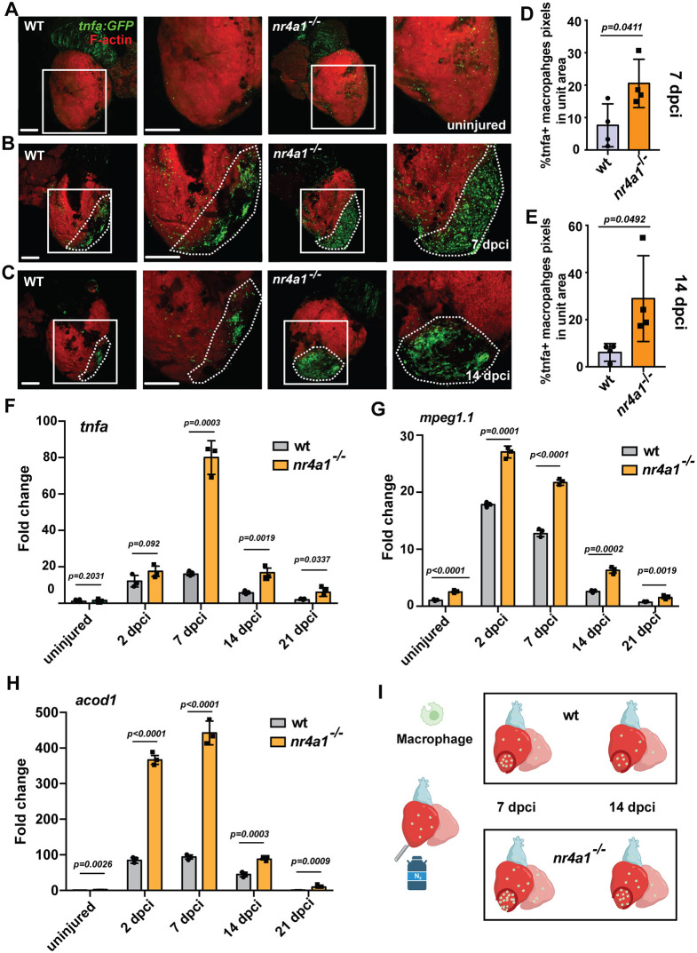
Abnormal accumulation of inflammatory macrophages in nr4a1 mutant. (A-C) Left: distribution of inflammatory macrophages in uninjured wild-type (WT) hearts and WT hearts at 7 and 14 dpci. Right: distribution of inflammatory macrophages in uninjured nr4a1 mutant hearts and nr4a1 mutant hearts at 7 and 14 dpci. Boxed regions in A-C show the approximate positions for quantification, and position of magnified images. Dashed lines show the wounded area. (D,E) Quantification of inflammatory macrophages in the unit area at 7 and 14 dpci. (F-H) qPCR results of inflammation-related genes tnfa, mpeg1.1 and acod1. All fold changes are calculated relative to the uninjured WT group in each panel. (I) Schematic showing inflammatory macrophage distribution in WT and nr4a1 mutant. Created in BioRender by Feng, D., 2025. https://BioRender.com/ynn17bt. This figure was sublicensed under CC-BY 4.0 terms. P-values<0.05 were considered statistically significant (two-tailed unpaired t-test, D,E; two-way ANOVA with Sidak test for multiple comparison correction and two-tailed unpaired t-test, F-H). Points in D and E show the individual samples. Data are mean±s.e.m. Scale bars: 275 µm.

Acknowledgments
This image is the copyrighted work of the attributed author or publisher, and ZFIN has permission only to display this image to its users. Additional permissions should be obtained from the applicable author or publisher of the image. Full text @ Development