
Image
|
Figure Caption
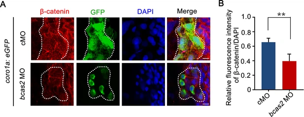
Fig. 3 - Supplemental 4 Knockdown of bcas2 significantly reduces nuclear β-catenin in the primitive myeloid cells. (A) Immunofluorescence staining of β-catenin in Tg(coro1a:GFP) embryos at 17 hpf. The embryos were injected with 8 ng of the indicated MOs at the one-cell stage and then collected for immunofluorescence staining. The dotted lines refer to the GFP-positive primitive myeloid cells. The relative fluorescence intensity of β-catenin was quantified in (B) (n=6). **p<0.01 (Student’s t-test). Scale bars, 10 μm (A).
Acknowledgments
This image is the copyrighted work of the attributed author or publisher, and
ZFIN has permission only to display this image to its users.
Additional permissions should be obtained from the applicable author or publisher of the image.
Full text @ Elife