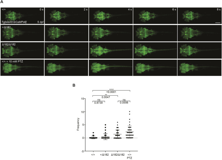
slc25a22aΔ182/Δ182 larvae exhibit elevated Ca2+ propagation from the midbrain to the spinal cord. (A) Tg(elavl3:Gal4,UAS:GCaMP6s) embryos at 5 dpf expressing GCaMP6s, a Ca2+ indicator, in mature neurons were subjected to time-lapse imaging for 20 min. WT larvae were exposed to 10 mM pentylenetetrazol (PTZ) for 30 min before the imaging. Scale bar: 300 μm. (B) Frequency of Ca2+ propagation from the midbrain to the spinal cord was measured for 20 min. Data are presented as mean±s.d. ns, not significant; **P<0.01 and ****P<0.0001 by one-way ANOVA with Tukey's HSD post hoc test (slc25a22a+/+, n=39; slc25a22a+/Δ182, n=40; slc25a22aΔ182/Δ182, n=42; WT+PTZ, n=40). Of note, the experiments shown in A and B were performed simultaneously with those in Fig. 6E,F using shared +/+ and Δ182/Δ182 groups.
This image is the copyrighted work of the attributed author or publisher, and
ZFIN has permission only to display this image to its users.
Additional permissions should be obtained from the applicable author or publisher of the image.
Full text @ Dis. Model. Mech.
Your Input Welcome
Thank you for submitting comments. Your input has been emailed to ZFIN curators who may contact you if
additional information is required.
Oops. Something went wrong. Please try again later.