IMAGE

Fig. 5

ID
ZDB-IMAGE-250609-81
Source
Figures for Eski et al., 2025
Image
Figure Caption

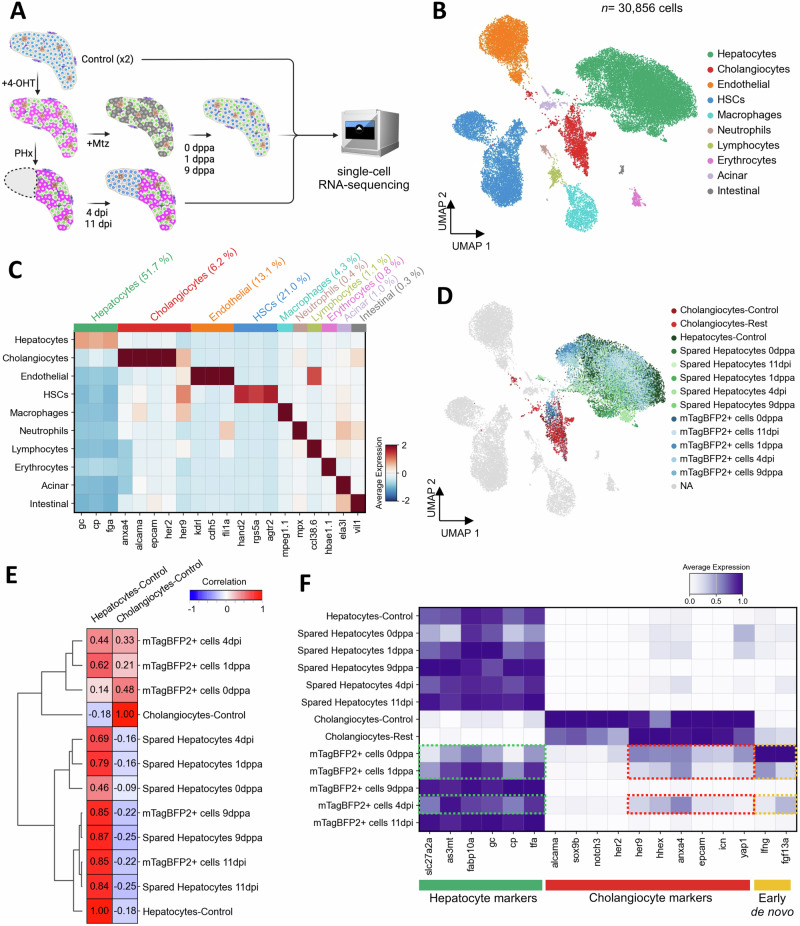
Fig. 5 De novo hepatocytes display transcriptional convergence with uninjured hepatocytes.

A Schematic representation of the single-cell RNA-seq approach to investigate the cellular and transcriptional landscape in basal condition (control-uninjured, N = 2) and during regeneration after partial ablation (0,1,9 dppa) and partial hepatectomy (4, 11 dpi). B UMAP visualization of cell types detected in zebrafish larval liver, combining data from injured and uninjured liver conditions. C Heatmap displaying the average scaled expression of selected markers across different cell types. D UMAP visualization focusing on specific injured conditions, highlighting mTagBFP2+ cells and spared hepatocytes (0,1,9 dppa and 4,11 dpi), control hepatocytes, control cholangiocytes, and cholangiocytes in injured conditions (cholangiocytes-rest). The remaining clusters are shown in gray. E Correlation matrix of hierarchically clustered mTagBFP2+ and spared hepatocytes compared to control hepatocytes and cholangiocytes. F Heatmap showing the normalized expression of hepatocyte and cholangiocyte-specific marker genes. Expression of selected genes that are detected in mTagBFP2+ cells at 0 dppa, 1 dppa and 4 dpi are outlined (dashed rectangles).

Acknowledgments
This image is the copyrighted work of the attributed author or publisher, and ZFIN has permission only to display this image to its users. Additional permissions should be obtained from the applicable author or publisher of the image. Full text @ Nat. Commun.