IMAGE

Figure 1

ID
ZDB-IMAGE-250607-4
Source
Figures for Wragg et al., 2025
Image
Figure Caption

Figure 1

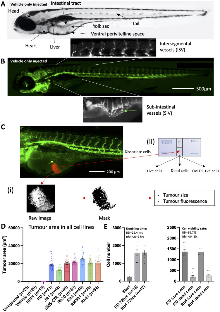
RMS tumour cells are viable and proliferate in larval zebrafish hosts. (A), Brightfield image of a 120 hpf zebrafish embryo highlighting key anatomical features, relevant to xenograft creation. (B), Matched fluorescent image illustrating the marking of developing blood vessels by the flk1:GFP transgene (green). The sub-intestinal vessel (SIV) and intersegmental vessel (ISV) beds are highlighted. (C), Schematic illustrating the techniques used to measure tumour burden. An image of a 120 hpf (70 hpi) zebrafish xenograft in flk1-GFP background is shown with blood vessels shown in green and the RMS tumour in red. Tumour induced reconfiguration of the SIV forming neo-vessel sprouts is marked (yellow arrow). (i) tumour size and fluorescence are assessed through FIJI image processing and analysis and (ii) tumour cell number and viability are assessed by excision of the tumour and dissociation, followed by trypan blue staining and cell counting on a haemocytometer. (D), Tumour area was calculated from fluorescent images as described in C, for 7 RMS cell lines alongside human foreskin fibroblasts (HFF1) as well as un-injected and vehicle (collagen) only injected zebrafish embryos. Data are presented as mean values +/- SEM. Data from 5 independent experiments for each line, with the exception of RD and HFF1 where n=6 and 3, respectively. (E), Bar chart showing total (left) and live/dead (right) cell number counts from excised RD and Rh4 tumours, measured at the end of the xenograft experiment (70 hpi) and compared to the initial injected bolus of 250 cells. Doubling time and cell viability rate are shown. Data from 2 independent experiments. n = number of xenografts.

Acknowledgments
This image is the copyrighted work of the attributed author or publisher, and ZFIN has permission only to display this image to its users. Additional permissions should be obtained from the applicable author or publisher of the image. Full text @ Front Oncol