
Image
|
Figure Caption
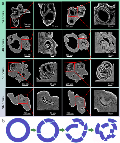
Fig. 6 High-resolution visualization of UBSEs in zebrafish intestines after 24, 48, 72, and 96 h of feeding. (a) The shells of UBSEs were found to gradually rupture, collapse, and shatter over time. Some UBSE shells still retained in the intestine even after 96 h of feeding. The slices were from the same zebrafish at each time point. (b) Schematic of the morphological changes of UBSE shells over time in the intestine. The UBSE shells gradually became fragmented.
Acknowledgments
This image is the copyrighted work of the attributed author or publisher, and
ZFIN has permission only to display this image to its users.
Additional permissions should be obtained from the applicable author or publisher of the image.
Full text @ Microsc. Res. Tech.