
|
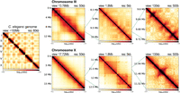
Fig. 1 3D organization of the C. elegans genome at different scales. Leftmost panel shows the Hi-C contact matrix of the entire genome. The right three columns show an example of an autosome (top) and X Chromosome (bottom) at different scales. At the chromosome-wide scale (first column), autosomes show clear separation between the two interacting flanking arms and the center, whereas the X Chromosome is more uniform. At the megabase scale (second column), autosomes show checkerboard pattern, whereas X Chromosome additionally harbors TADs. At the kilobase scale (last column), both autosomes and X Chromosome show fountains that appear as protruding 3D DNA contacts from the main diagonal.