
|
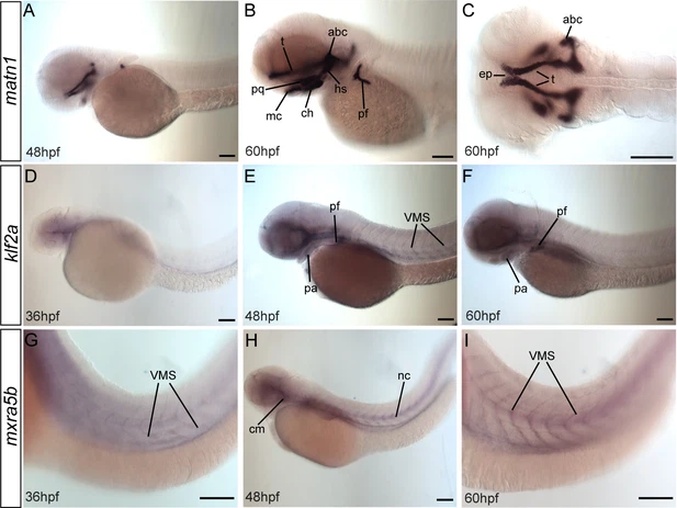
Fig. 2 - Supplement 1 matn1, klf2a, and mxra5b are expressed in musculoskeletal tissues of developing embryos. Lateral (A, B, D–I) and ventral (C) views of embryos showing expression of matn1 (A–C), klf2a (D–F), and mxra5b (G, H). (A–C) 48 hpf embryos show matn1 expression in cartilage progenitors at 48 hpf and in differentiated cartilages (and associated tenocytes) at 60 hpf (B, C). Lateral views of 36 hpf (D), 48 hpf (E), and 60 hpf (F) embryos show klf2a expression in pharyngeal mesenchyme (D), skeletal progenitors and in tenocytes along VMS (E, F). Lateral views of 36 hpf (G), 48 hpf (H), and 60 hpf (I) embryos show mxra5b expression in tenocytes along horizontal myosepta (HMS) along the notochord and VMS. Scale bars = 100 μm. Abbreviations: abc = anterior basicranial commissure, ch = ceratohyal cartilage, ep = ethmoid plate, hs = hyosymplectic cartilage, mc = meckel’s cartilage, nc = notochord, pf = pectoral fin, pq = palatoquadrate cartilage, sb = somite boundaries, t = trabeculae cartilage.