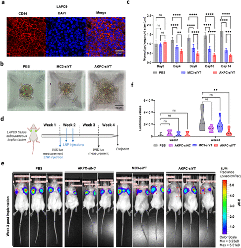
Fig. 7 Antitumor effect of AKPC-siYT on prostate cancer PDX-derived organoids and in vivo tumor growth. a, Prostate cancer PDX model (LAPC9) exhibited high CD44 protein levels (CD44 in red, nuclei marked by DAPI in blue) by immunofluorescence staining. b, Morphology of LAPC9 PDX-derived organoids following treatment with PBS, MC3-siYT, and AKPC-siYT (siRNA, 2 μg/mL) for 14 days. Scale bar, 50 μm. c, Organoid size of each treatment group (PBS, MC3-siYT, and AKPC-siYT) was measured at different time points (day 0, 4, 8, 10, and 14). Two-way ANOVA multiple comparison was used to determine the significance of the comparisons of data (*p < 0.05; **p < 0.01; ***p < 0.001; ****p < 0.0001). In all panels, error bars represent mean ± s.d. (n = 3) d, Schematic representation of in vivo tumor growth kinetics of LAPC9 PDX following LNP treatment. Bioluminescent LAPC9-copGFP-CBR tumor tissues were subcutaneously (s.c.) implanted in CB17 SCID mice at day 0. Following a lag period of 1 week, mice were subjected to one-week LNP treatment (Days 7, 9, and 11 of week 2). LNPs were sc injected at the tumor-adjacent area (siRNA, 10 μg/tumor, n = 3/group). Intravital imaging (IVIS-CT) was used to record tumor dynamics based on stable bioluminescence expression of the LAPC9 tumor cells weekly for 3 consecutive weeks. At the endpoint (week 4), IVIS-CT, tumor collection, and body weight measurement were done. e, Bioluminescence images of LAPC9 PDX tumors showing individual tumor areas (n = 3/LNP treatment group) at the endpoint. f, Violin plot of in vivo tumor growth based on average bioluminescence radiance of individual tumors at endpoint (day 28) (n = 3 animals/group × 2 tumors). Two-way ANOVA, ŠídÁk’s multiple comparisons test was used to determine the significance of the comparisons of data indicated in d (*p < 0.05; **p < 0.01; ***p < 0.001; ****p < 0.0001). In all panels, error bars represent mean ± s.d.
Image
Figure Caption
Acknowledgments
This image is the copyrighted work of the attributed author or publisher, and
ZFIN has permission only to display this image to its users.
Additional permissions should be obtained from the applicable author or publisher of the image.
Full text @ ACS Nano

