
|
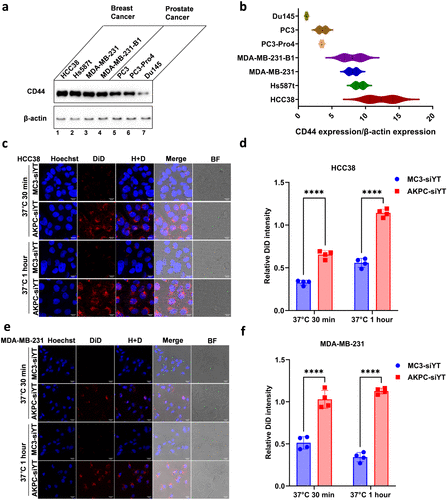
Fig. 2 Evaluation of AKPC-siYT targeting breast cancer cells in vitro. a, Western blot images of CD44 expression in different cell lines. CD44 and β-actin mouse primary antibodies were used to detect protein expression. b, Quantification of CD44 expression to β-actin in different cell lines. c,e, Confocal microscopic images of cellular internalization of LNPs in HCC38 and MDA-MB-231 cells at 37 °C after 30 min and 1 h of incubation. 0.5 mol % DiD was added to the lipids and served as the fluorescent dye. Scale bar represents 20 μm. d,f, The DiD fluorescence intensity was normalized to Hoechst for the uptake quantification of LNPs by HCC38 and MDA-MB-231 cells. A two-way ANOVA multiple comparison was used to determine the significance of data indicated in d and f (*p < 0.05; **p < 0.01; ***p < 0.001; ****p < 0.0001). In all panels, error bars represent mean ± s.d. (n = 3).