
|
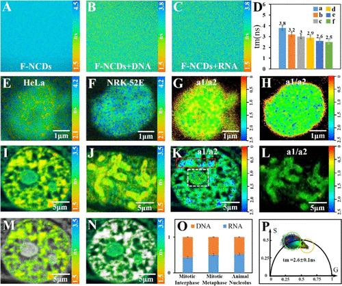
Fig. 3 (A–C) FLIM imaging of F-NCDs in pure solution, F-NCDs bound to DNA, and F-NCDs bound to RNA in mixed solution. (D) Fluorescence lifetime change of F-NCDs binding to different concentrations of DNA and RNA (a: F-NCDs; b: F-NCDs+100%RNA; c: F-NCDs + 75%RNA + 25%DNA; d: F-NCDs + 50%RNA + 50%DNA; e: F-NCDs + 25%RNA + 75%DNA; f: F-NCDs + 100%DNA). (E, F) FLIM images of nucleolus in HeLa and NRK-52E cells stained with F-NCDs. Co-localization results of F-NCDs with the commercial dye Hoechst 33342 showed that F-NCDs targeted the nucleolus of HeLa and NRK-52E cells (Figure S14) and that the F-NCDs had better photostability (Figure S15). (G, H) a1/a2 ratio of (E, F), respectively. Color-coded by the a1/a2 ratio. (I, J) FLIM image of a single plant cell stained with F-NCDs in interphase and metaphase. (K, L) a1/a2 ratio of (I, J). (M, N) FLIM images of DNA and RNA separated by a phasor plot. (O) Comparison of DNA and RNA ratios in different species and mitotic stages. (P) Phasor plot corresponding to (I). All a1/a2 ratio images were denoised.