IMAGE

Figure 3

ID
ZDB-IMAGE-250419-15
Source
Figures for Su et al., 2025
Image
Figure Caption

Figure 3

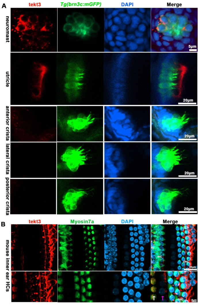
TEKT3/Tekt3 is present in some HCs. (A) Neuromast and utricular HCs pf zebrafish larvae at 5 dpf are positive for anti-Tekt3 antibody staining while the crista HCs are negative. (B) TEKT3 is present in mature mouse cochlear HCs. The left row of cells are inner HCs (IHC, pointed by a white arrow) and the right three rows are outer HCs (pillar cell, pointed by a pink arrow). Note: The saccular and crista HCs are always negative.

Acknowledgments
This image is the copyrighted work of the attributed author or publisher, and ZFIN has permission only to display this image to its users. Additional permissions should be obtained from the applicable author or publisher of the image. Full text @ Int. J. Mol. Sci.