
Fig. 3
- ID
- ZDB-IMAGE-250416-80
- Publication
- Yang et al., 2025 - Crip2 affects vascular development by fine-tuning endothelial cell aggregation and proliferation
- All Figures
- Figures for Yang et al., 2025
|
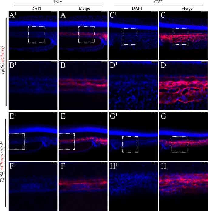
Fig. 3 Loss of CRIP2 leads to aggregation of vascular endothelial cells. No endothelial cell aggregation was observed in the PCV (A, B) or CVP (C, D) of Tg(flk: mCherry) zebrafish embryos at 48 hpf. Aggregation of vascular endothelial cells was observed in the PCV (E, F) and CVP (G, H) of Tg(flk: mCherry); crip2−/− homozygous mutant embryos at 48 hpf. The embryos were stained with DAPI for nuclear visualization, with the left panel showing the embryonic trunk and the right panel showing the embryonic tail bud. The white dashed boxes indicate the areas that are magnified in the corresponding enlarged images located below in the figures. PCV: posterior cardinal vein; CVP: caudal vein plexus