
|
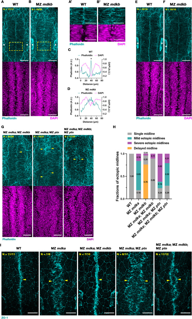
Fig. 3 A mutation in mdkb rescues midline defects in MZ mdka mutants. (A-B) Representative MIP images of Phalloidin (cyan) and DAPI (magenta) stained WT and MZ mdkb mutant embryos at 17 hpf. Distinctive F-actin accumulation in medial region of WT (A), in contrast to MZ mdkb mutants (B). Nuclei distribution was similar in WT and MZ mdkb mutants (lower images). Positions of otic vesicles are labelled by white asterisks. Yellow dashed boxes indicate regions shown with higher magnification in (A′-B′). Scale bars = 50 μm (A-B) and 30 μm (A′-B′). (C-D) Quantification of mean fluorescent intensity of Phalloidin (cyan) and DAPI (magenta) along left-right axis of (A′-B′). Asterisks show the accumulation of Phalloidin-stained F-actin. (E-F) At 18 hpf, distinctive F-actin accumulation was found in both WT and MZ mdkb mutant embryos. The positions of otic vesicles are indicated by asterisks. Scale bars = 50 μm. (G) Representative MIP images of Phalloidin stained F-actin (cyan) and DAPI stained nuclei (magenta) in rhombomere region of WT, MZ mdka; MZ mdkb, MZ mdka; MZ ptn double mutant and MZ mdka; MZ mdkb; MZ ptn triple mutant embryos at 17 hpf. Yellow arrowheads indicate F-actin accumulation at a single midline in WT and MZ mdka; MZ mdkb mutants and at ectopic midlines in MZ mdka; MZ ptn double and MZ mdka; MZ mdkb; MZ ptn triple mutants. Scale bar = 50 μm. (H) Categorization of midline phenotypes in WT, MZ mdka, MZ mdkb, MZ mdka; MZ mdkb, MZ mdka; MZ ptn and MZ mdka; MZ mdkb; MZ ptn mutants. Data of WT and MZ mdka was also shown in Fig. 2H–J. (I) Representative MIP dorsal views after ZO-1 (cyan) staining in WT, MZ mdka, MZ mdka; MZ mdkb, MZ mdka; MZ ptn and MZ mdka; MZ mdkb; MZ ptn mutants at 17 hpf. Accumulation of ZO-1 is indicated by yellow arrowheads. Scale bar = 50 μm. (For interpretation of the references to colour in this figure legend, the reader is referred to the Web version of this article.)
Reprinted from Developmental Biology, 521, Le, Y., Rajasekhar, K., Loo, T.Y.J., Saunders, T.E., Wohland, T., Winkler, C., Midkine-a interacts with Ptprz1b to regulate neural plate convergence and midline formation in the developing zebrafish hindbrain, 52-74, Copyright (2025) with permission from Elsevier. Full text @ Dev. Biol.