
Image
|
Figure Caption
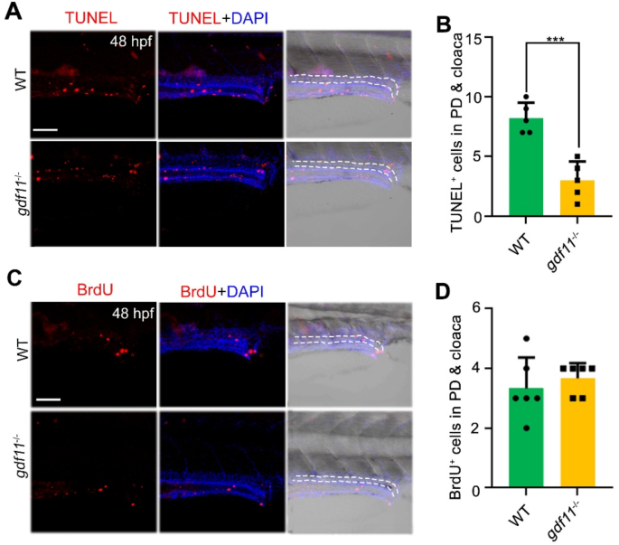
Fig. 4 The apoptosis of pronephric tubules and cloaca was significantly decreased upon gdf11 deletion. (A,B) TUNEL assay showing decreased apoptosis in cells of the developing pronephric duct and cloaca in gdf11 mutants. The position marked by the dotted line in the figure is the pronephros and the cloaca. Scale bar: 50 μm. (C,D) Cell proliferation, indicated by BrdU staining, remains unchanged in both WT and gdf11 mutants. The position marked by the dotted line in the figure is the pronephros and the cloaca. Scale bar: 50 μm.
Acknowledgments
This image is the copyrighted work of the attributed author or publisher, and
ZFIN has permission only to display this image to its users.
Additional permissions should be obtained from the applicable author or publisher of the image.
Full text @ Sci. Rep.