
|
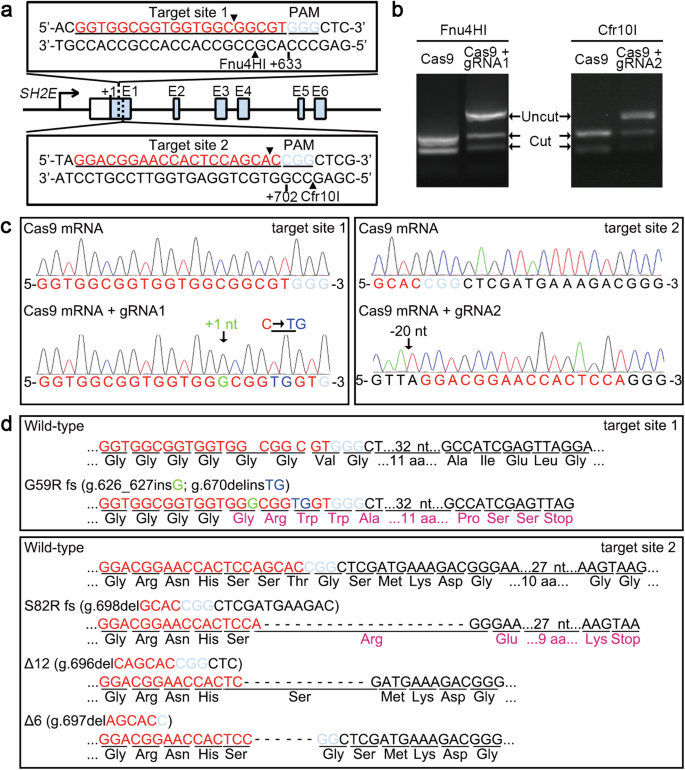
Fig. 2 a Schematic structure of CRISPR targeting on zebrafish SH2E gene. Two target sites of CRISPR were designed in exon 1 to induce SH2E deficiency. The target sites of two restriction enzymes, Fnu4H I and Cfr10 I, are indicated with black triangles. +633 and +702 indicate the positions of PAM sequences. b The genotypes of embryos injected with Cas9 mRNA alone or with gRNA1/2 were confirmed by enzyme digestion using Fnu4H I and Cfr10 I, respectively. In each test, 10 embryos injected were randomly selected and pooled as one sample. c The mutated genomic sequences were confirmed by sequencing. d G59R fs and S82R fs are two types of mutants with frame shifts leading to early termination of protein translation. Δ12 and Δ6 are two types of mutants without frame shifts. The altered amino acid sequences are labeled in purple.