
Image
|
Figure Caption
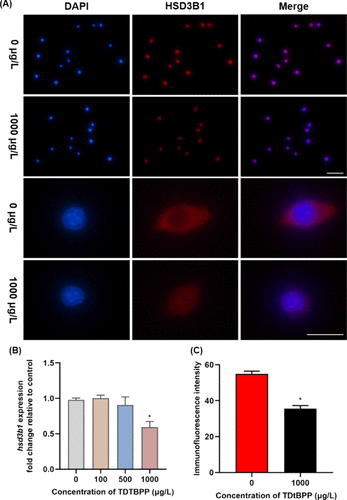
Fig. 5 Figure 5. Immunofluorescence staining (A) and fluorescence signal intensity (C) of HSD3B1 in Leydig cells in 0 and 1000 μg/L TDtBPP exposure groups. Blue indicates the nucleus, and red indicates the HSD3B1 protein. Scale bar is 50 μm above and 100 μm below. Expression of hsd3b1 (B, n = 4) in Leydig cells after exposure to 0, 100, 500, or 1000 μg/L TDtBPP. *p < 0.05 was considered to indicate a significant difference.
Acknowledgments
This image is the copyrighted work of the attributed author or publisher, and
ZFIN has permission only to display this image to its users.
Additional permissions should be obtained from the applicable author or publisher of the image.
Full text @ Env. Sci. Tech.