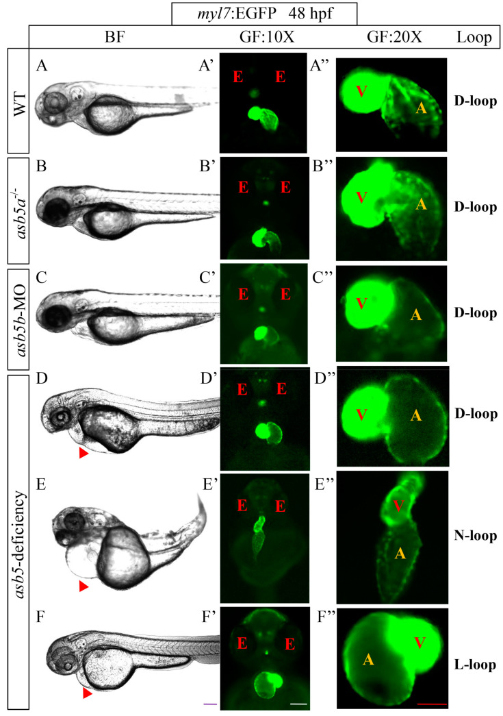
IMAGE

Figure 1

ID
ZDB-IMAGE-250329-86
Source
Figures for Zhou et al., 2025
Image
Figure Caption

Figure 1

The impact of asb5a/asb5b deficiency on L-R asymmetry in cardiac development. (AF) Representative images of the pericardial cavity in various groups at 48 hpf during embryonic development; scale bar = 200 µm (side view). (A′F′) Representative images of heart positioning in different zebrafish groups at 48 hpf under the myl7:EGFP background; scale bar = 100 µm (belly up view); (A″F″) Magnified images of the atria and ventricles in various zebrafish groups at 48 hpf, also under the myl7:EGFP background, scale bar = 50 µm (belly up view). The red triangular arrows indicate areas of enlargement in the cardiac pericardial cavity. BF: bright field; GF: green fluorescence; 10X: field of view under a 10× microscope; 20X: field of view under a 20× microscope; red E: eyes; red V: ventricle; yellow A: atrium; D-loop: dextral looping; N-loop: no looping; L-loop: levo-looping.

Acknowledgments
This image is the copyrighted work of the attributed author or publisher, and ZFIN has permission only to display this image to its users. Additional permissions should be obtained from the applicable author or publisher of the image. Full text @ Int. J. Mol. Sci.