IMAGE

Fig 4

ID
ZDB-IMAGE-250321-13
Source
Figures for Nonarath et al., 2025
Image
Figure Caption

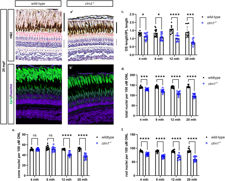
Fig 4 clrn1-/- zebrafish present with age-dependent shortening of the rod outer segments and thinning of the outer nuclear layer.

Hematoxylin and eosin-stained paraffin section from (a) 20 mpf wild-type and (a’) clrn1-/- zebrafish. Staining for Zpr3+ (Rhodopsin and green opsin), primarily marking rod photoreceptor outer segments (green) on paraffin sections from (b) 20 mpf wild-type and (b’) clrn1-/- zebrafish. (c) Quantification of rod outer segments (OS) length at 4, 8, 12, and 20 mpf. (d) Quantification of total photoreceptors per 100 µm of ONL length at 4, 8, 12, and 20 mpf. Analysis of (e) rod and (f) cone photoreceptors in wild-type and clrn1-/- zebrafish at 4, 8, 12, and 20 mpf. (a-b’) The black bars in (a’) denote the location of the rod OS, and the grey bar notes the outer nuclear layer. (ns, not significant = p>0.05, *p<0.05, **p<0.01, ***p<0.005, ****p<0.001; One-way ANOVA). (n=12-15 for each genotype per time-point).

Acknowledgments
This image is the copyrighted work of the attributed author or publisher, and ZFIN has permission only to display this image to its users. Additional permissions should be obtained from the applicable author or publisher of the image. Full text @ PLoS Genet.