
Image
|
Figure Caption
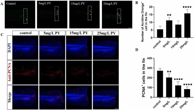
Fig. 3 Pyrazosulfuron-ethyl influenced celluar proliferation and apoptosis in regenerative fins. (A). 24 hpa AO staining in caudal fin. (B). Statistics of AO labeled apoptotic cell data in zebrafish larvae at 24 hpa (C). 24 hpa PCNA antibody staining in caudal fin area. (D). Statistics of the number of PCNA+ labeled proliferative cells in the caudal fin at 24 hpa. (Data represent mean ± S.D. ns>0.05, ** p < 0.01, **** p < 0.0001. ns is not labelled in the graph. Scale bar: (A) 100 μm, (C,D) 75 μm).
Acknowledgments
This image is the copyrighted work of the attributed author or publisher, and
ZFIN has permission only to display this image to its users.
Additional permissions should be obtained from the applicable author or publisher of the image.
Full text @ Ecotoxicol. Environ. Saf.