IMAGE

Fig 6

ID
ZDB-IMAGE-250313-7
Source
Figures for Sarich et al., 2025
Image
Figure Caption

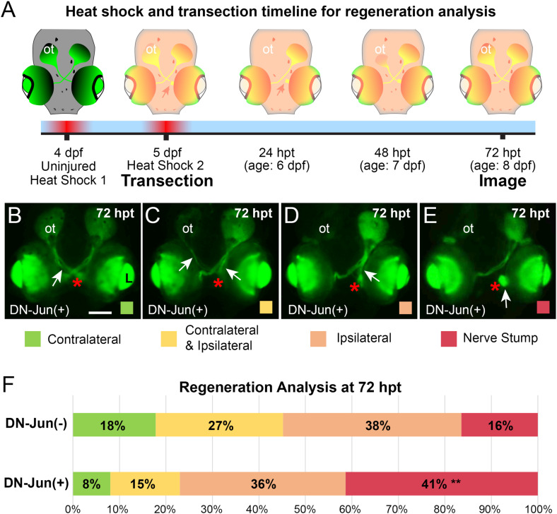
Fig 6 Induction of DN-Jun diminishes capacity for optic nerve regeneration.

A. Experimental workflow. Left optic nerves of Tg(isl2b:GFP) X Tg(mke15Tg) progeny were transected after the second heat shock at 5 dpf then allowed to regenerate through 72 hpt (age: 8 dpf) when they were live imaged and analyzed. (B–E) Representative live fluorescent images of 72 hpt (age: 8 dpf) DN-Jun expressing larvae and the four potential regeneration outcomes after optic nerve transection. Red asterisks =  site of injury. B. Primarily contralateral axon regeneration, visible GFP labeled axons navigate to the opposite side’s optic tectum, white arrow. C. Contralateral and ipsilateral axon regrowth were both visible, and it was not possible to determine a dominant path, white arrows. D. Visible regenerating axons were primarily directed ipsilaterally, white arrow. E. No visible regeneration occurred; a nerve stump was present, white arrow. F. 100% stacked bar graph of regeneration progress in DN-Jun(-) and DN-Jun(+) fish. Color code indicates the primary axon growth trajectory identified. Green =  contralateral axon growth. Yellow =  both contralateral and ipsilateral axon growth. Orange =  ipsilateral axon growth. Red =  nerve stump, with no visible axon growth. Chi-square analysis was used to determine the significance between DN-Jun(-) and DN-Jun(+) regeneration phenotypes. The nerve stump phenotype was significantly increased in DN-Jun(+), ** P =  0.0025. Graph represents five biological replicates, DN-Jun(-) (negative for DN-Jun after heat shock) n =  73; DN-Jun(+) (positive for DN-Jun after heat shock) n =  87. ot =  optic tectum, L =  lens. Scale bar =  100 µm.

Acknowledgments
This image is the copyrighted work of the attributed author or publisher, and ZFIN has permission only to display this image to its users. Additional permissions should be obtained from the applicable author or publisher of the image. Full text @ PLoS One