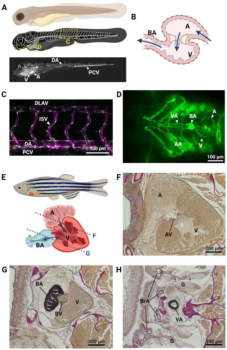
IMAGE

Figure 1

ID
ZDB-IMAGE-250311-44
Source
Figures for Prendergast et al., 2025
Image
Figure Caption

Figure 1

The zebrafish cardiovascular system. Representation of key elements of the zebrafish cardiovascular anatomy in embryonic/larval (A–D) and adult (E–H) stages. (A) Schematic overview of the circulatory system in a zebrafish embryo, showing the location of images in panels C and D. bottom: angiogram of a 5 days post fertilization (dpf) Tg(gata1:dsRed) zebrafish embryo, showing the trajectory of fluorescent erythrocytes throughout the circulation. (B) Schematic representation of the zebrafish embryo heart, with indication of blood flow direction (arrows). (C) Lateral view of trunk blood vessels in a 2dpf Tg(kdrl:ras-mCherry;fli1a:nls-gfp) zebrafish embryo, with endothelial cell membranes in magenta and endothelial cell nuclei in green. (D) Ventral view of the head of a Tg(kdrl:egfp) 4 dpf zebrafish larva, showing the heart and outflow tract. (E) Schematic overview of the adult zebrafish heart. The approximate planes of the cardiac sections in panels (F) and (G) are indicated with a dotted line. (F–H) Resorcin-fuchsin stain of elastin fibers (dark purple to black) in sections of a 9-month-old male zebrafish. (F,G) cardiac chambers and valves, (H) cross-section of the ventral aorta. AA, aortic arches; A, atrium; AV, atrioventricular valve; BA, bulbus arteriosus; BrA, branchial arteries; BV, bulboventricular valve; DA, dorsal aorta; DLAV, dorsal longitudinal anostomotic vessels; G, gills; ISV, intersegmental vessel; PCV, posterior caudal vein; V, ventricle; VA, ventral aorta. Created in BioRender. Sips, P. (2025) https://BioRender.com/z89q283.

Acknowledgments
This image is the copyrighted work of the attributed author or publisher, and ZFIN has permission only to display this image to its users. Additional permissions should be obtained from the applicable author or publisher of the image. Full text @ Front Cardiovasc Med