IMAGE

Fig. 1.

ID
ZDB-IMAGE-250311-10
Source
Figures for Varady et al., 2025
Image
Figure Caption

Fig. 1.

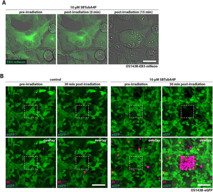
Local illumination of SBTubA4P ablates cancer cells with spatial control in vitro. (A) Repeated SBTubA4P illumination causes blebbing, contraction and swelling of cancer cells. OS143B-EB3-mNeon cells were incubated with 10 µM SBTubA4P for 1 h. Illumination was performed by repeated laser scanning with the 405 nm UV laser diode of a confocal microscope (Leica SP8 WLL) for 3 min (∼15 s between scans). Standstills from the subsequent timelapse (see Movie 2) show the onset of contraction and blebbing of cells (0 min timepoint) followed by swelling and loss of fluorescence (15 min timepoint). Representative confocal images from three independent experiments. Scale bar: 25 µm. (B) Spatially confined SBTubA4P illumination induces localized cell death in OS143B cells. OS143B-eGFP cells were incubated with 10 µM SBTubA4P for 1 h before illumination or left untreated (control). Propidium iodide (PI) solution was added to the medium before imaging. UV irradiation by repeated scanning on a confocal microscope was performed for 5 min in an area of 95×95 µm, depicted by the dashed line squares. PI staining and loss of eGFP fluorescence indicate cell death (see Movies 3 and 4). Representative confocal images (n=3 per condition). Scale bars: 100 µm.

Acknowledgments
This image is the copyrighted work of the attributed author or publisher, and ZFIN has permission only to display this image to its users. Additional permissions should be obtained from the applicable author or publisher of the image. Full text @ Dis. Model. Mech.