IMAGE

Fig. 5

ID
ZDB-IMAGE-250307-55
Source
Figures for Hu et al., 2025
Image
Figure Caption

Fig. 5

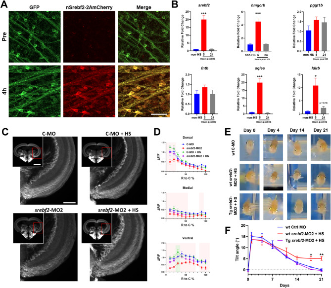
Conditional expression of constitutively active srebf2 rescues loss-of-function but does not accelerate regeneration. (A) mCherry expression in whole mount Tg(hsp70l: nSrebf2-2AmCherry, cmlc: GFP) zebrafish retina GCL before heat shock (Pre) and 4-hours post heat shock (4 h). (B) Expression of selected mev/chol synthesis pathway genes in post heat shock retina measured by qRT-PCR by one-way ANOVA with Bonferroni post hoc test, n = 4 for each group. (C) Representative images of axon regeneration into the optic tectum after treatment with control MO (C-MO) or srebf2-MO2 with and without heat shock rescue (+ HS) by Tg(hsp70l: nSrebf2-2AmCherry, cmlc: GFP). (D) Quantification of regeneration into the optic tectum. Red boxes represent p < 0.05 in C-MO vs. srebf2-MO2 group at the corresponding rostral to the caudal region; green boxes represent p < 0.05 in C-MO vs. C-MO + HS by two-way ANOVA with Fisher’s LSD post hoc test, no other comparisons were statistically significant, n = 5–6 for each group. (E) Representative images of the DLR on various days post nerve injury with C-MO, srebf2-MO2, and srebf2-MO2 plus transgenic heat shock rescue. (F) Quantification of the DLR in each treatment group over time by two-way ANOVA with Bonferroni post hoc test, n = 5–6 for each group. + HS indicates daily heat shock treatment starting at day 0. Scale bar in A = 50 μm. Scale bar in C = 500 μm and 200 μm. * p < 0.05, ** p < 0.01, *** p < 0.001

Acknowledgments
This image is the copyrighted work of the attributed author or publisher, and ZFIN has permission only to display this image to its users. Additional permissions should be obtained from the applicable author or publisher of the image. Full text @ Mol. Neurodegener.