IMAGE

Fig. 3

ID
ZDB-IMAGE-250307-53
Source
Figures for Hu et al., 2025
Image
Figure Caption

Fig. 3

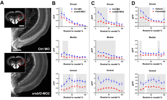
Srebf2 loss-of-function inhibits axon regeneration in the optic tectum at 7 dpi. (A) Representative images of tectum and higher magnification of medial-ventral tectum of vehicle or fatostatin treated zebrafish. (B) Quantification of tectum regeneration at dorsal, medial, and ventral region after srebf2-MO1 treatment. (C) Quantification of tectum regeneration at dorsal, medial, and ventral region after srebf2-MO2 treatment. (D) Quantification of tectum regeneration at dorsal, medial, and ventral region post fatostatin treatment. Grey boxes indicate p < 0.05 compared with Ctrl MO or Vehicle at the corresponding rostral to caudal distance by two-way ANOVA with Fisher’s LSD post hoc test. Scale bar = 500 and 200 μm, n = 5–6 for each group

Acknowledgments
This image is the copyrighted work of the attributed author or publisher, and ZFIN has permission only to display this image to its users. Additional permissions should be obtained from the applicable author or publisher of the image. Full text @ Mol. Neurodegener.