
Image
|
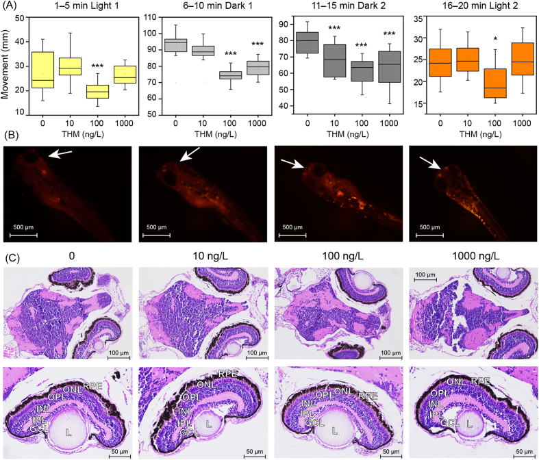
Figure Caption
Fig. 1
(A) Photomotor response of zebrafish larvae at 120 hpf. (B) Fluorescent microscopic images of the larvae at 120 hpf, with white arrow indicating fluorescence around the eyes. (C) Histological changes in the brain and retinal tissues of zebrafish larvae at 120 hpf. GCL, ganglion cell layer; INL, inner nuclear layer; IPL, inner plexiform layer; L, lens; ONL, outer nuclear cell; OPL, outer plexiform layer; REP, retinal pigmented epithelium.
Acknowledgments
This image is the copyrighted work of the attributed author or publisher, and
ZFIN has permission only to display this image to its users.
Additional permissions should be obtained from the applicable author or publisher of the image.
Full text @ Eco Environ Health